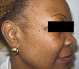
Dermatosis papulosa nigra, also known as DPN, are small, soft brown papules that may occur on the face and neck of patients of African, Latin, Indian, or Asian descent. While they may not reach the size of their histologically similar seborrheic keratosis counterparts, the lesions do represent a sign of aging in darker skinned patients. However, the lesions can be safely, easily, and effectively removed.
Electrodesiccation with a hyfrecator or destruction with the KTP (532 nm) laser are my favorite methods for DPN removal. I prefer not to use curettage or cryotherapy because of the risk for dyspigmentation in darker skinned patients. Case reports of success with fractional photothermolyis (1,550 nm) and Nd:YAG lasers (1,064 nm) have been published.
 2009/Elsevier
Dermatosis papulosa nigra can be safely and effectively treated via electrodesiccation or with a KTP laser.
If electrodesiccation is performed, the application of topical anesthetic prior to the procedure helps make the patient more comfortable. For larger lesions, injection of 1% lidocaine with 1:100,000 epinephrine may be used.
Also, with electrodesiccation, conservative settings (0.6-2.0 W on the low setting) should be used; the lesions are desiccated using a blunt tip for a few seconds until they turn grayish.
Care is taken not to touch the surrounding skin. A sharp tip may be used with very small (less than 1 mm) lesions for more accurate precision. I wipe the tip from time to time with gauze to avoid char accumulation.
Larger or pedunculated lesions may be treated with electrodesiccation or snipped off with gradle scissors.
With the KTP laser, topical anesthesia is usually not required. I use a smaller spot size than the lesion itself to avoid targeting and potentially causing dyspigmentation of the surrounding skin.
A spot size of 1 mm is typically used, with 6-10 ms and 10-15 j/cm2. The laser tip is held approximately 1 cm away from the skin at a 90 degree angle. I start off with the lowest fluence and adjust it higher until the lesions turn grayish and a light popping sound is heard with the laser pulse.
A split-face study published in the American Journal of Dermatologic Surgery in 2009 showed that both electrodesiccation and KTP have comparable efficacy in removal of DPN. Without use of anesthetics, the KTP laser was preferred for patient comfort.
Immediately after treatment, patients can expect the treated lesions to become red and swollen - similar to insect bite reactions - for about an hour. Antibiotic ointment or aquaphor is applied to soothe the skin.
Patients are then told to leave the lesions alone, to avoid picking, and to avoid sun exposure. Patients are also advised to avoid alpha-hydroxy acids and other "anti-aging" products until healed. If the cheeks were treated, make-up (foundation, blush) may be applied in 3 to 4 days. Lesions typically fall off within a week.
If needed, repeat treatment may be performed in 2 to 4 weeks.
If you have any DPN removal tips, please feel free to share!
-Naissan Wesley, M.D.
Do you have questions about treating patients with darker skin? If so, send them to sknews@elsevier.com.